Home
/ Human Bone Anatomy Ribs : The Bones of the Thorax - the rib cage | Thorax, Anatomy ... / The ribs, together with the sternum form the rib cage, which protects the vital organs and serves as attachment sites for muscles.
Human Bone Anatomy Ribs : The Bones of the Thorax - the rib cage | Thorax, Anatomy ... / The ribs, together with the sternum form the rib cage, which protects the vital organs and serves as attachment sites for muscles.
Human Bone Anatomy Ribs : The Bones of the Thorax - the rib cage | Thorax, Anatomy ... / The ribs, together with the sternum form the rib cage, which protects the vital organs and serves as attachment sites for muscles.. Human body anatomy ribs intestines heart bones torso popsockets grip and stand for phones and tablets. Yet, the ribs and rib cage are also flexible enough to expand. Have you ever seen fossil remains of dinosaur and ancient human bones in textbooks, television, or in person at a museum? Human body skeleton system bone joints anatomy. What about the transition from molecules to man?
Ross and wilson has been a core text for students of anatomy and physiology. Human structure and functions in health. Your rib cage, for example, acts like a shield around your chest to protect important organs inside such as your lungs and heart. Human body anatomy ribs intestines heart bones torso popsockets grip and stand for phones and tablets. The study of bone cartilage:
Ribs: Anatomy, Types, Ossification & Clinical Significance ... from www.howtorelief.com Bone basics and bone anatomy. Later lessons will cover each of these bones in further detail. Bones are organs that produce red and white blood cells, store minerals, enable mobility, and provide structural support for the body. In in depth source to learn and see the anatomy of all the the axial skeleton consists of the central core of the skull, spine, and ribs whilst the appendicular is. Flexible connective tissue composed of collagen and elastin fibres. The vertebrae when we lift heavy weights; Vertebrae in the thoracic spine on the other hand are less mobile due to being joined by the ribs to the sternum. Human body skeleton system bone joints anatomy.
The costotransverse ligaments in human:
But this number may be increased by the development of a cervical or lumbar rib, or may be diminished to eleven. The key bones of the human body. From the anatomy of the human rib cage, we can tell that the human ribs bones have several parts: Costae) are the long curved bones which form the rib cage, part of the axial skeleton. Long bones, short bones, and flat bones. Yet, the ribs and rib cage are also flexible enough to expand. The ribs are curved, flat bones which form the majority of the thoracic cage. Photo human body bone joint pains anatomy (ribs). The hand bones are also known as carpel bones. Human body anatomy, body silhouette. In vertebrate anatomy, ribs (latin: Red bone marrow contains hematopoietic stem cells that produce two other types of stem cells: The relative quantity of these two kinds of tissue varies in different bones.
The key bones of the human body. .of human bones pdf, anatomy of human bones ppt, anatomy of the body muscles and bones, anatomy of the moving body a basic course in bones human anatomy, anatomy heart, anatomy pelvis, anatomy rib cage organs, anatomy ribs, anatomy sternum, anatomy xiphoid process, male. Lessons on the skeletal system (upper limb, lower limb, skull, vertebrae, rib, and sternum bones). Vintage anatomical drawing medical illustration , pelvis , hip skeleton book page , paper ephemera human anatomy. The costotransverse ligaments in human:
Rib cage bones human skeletal system anatomy Vector Image from cdn5.vectorstock.com Vintage anatomical drawing medical illustration , pelvis , hip skeleton book page , paper ephemera human anatomy. The wider section at each end of the bone is called the epiphysis (plural the two layers of compact bone and the interior spongy bone work together to protect the internal organs. Illustration of rib cage, demonstrating ribs and connection through cartilage to sternum. The first seven are connected behind with the vertebral column. Observation and analysis method for human bones chap. Your rib cage, for example, acts like a shield around your chest to protect important organs inside such as your lungs and heart. Individual bone models | human anatomy. Costae) are the long curved bones which form the rib cage, part of the axial skeleton.
The ribs are elastic arches of bone, which form a large part of the thoracic skeleton.
Have you ever seen fossil remains of dinosaur and ancient human bones in textbooks, television, or in person at a museum? .of human bones pdf, anatomy of human bones ppt, anatomy of the body muscles and bones, anatomy of the moving body a basic course in bones human anatomy, anatomy heart, anatomy pelvis, anatomy rib cage organs, anatomy ribs, anatomy sternum, anatomy xiphoid process, male. Lessons on the skeletal system (upper limb, lower limb, skull, vertebrae, rib, and sternum bones). Diagram of organs of the human body. Contributing to their role in protecting the internal thoracic organs. Bones are not inanimate rock like structures in the human body; They are extremely light, but highly resilient; The ribs are elastic arches of bone, which form a large part of the thoracic skeleton. If the outer layer of a cranial bone fractures, the. The ribs also bend a little as we breathe; It possesses also a certain degree the compact tissue is always placed on the exterior of the bone, the cancellous in the interior. The vertebrae when we lift heavy weights; Vertebrae in the thoracic spine on the other hand are less mobile due to being joined by the ribs to the sternum.
This section is only a brief overview and introduction. In in depth source to learn and see the anatomy of all the the axial skeleton consists of the central core of the skull, spine, and ribs whilst the appendicular is. Bones are not inanimate rock like structures in the human body; Individual bone models | human anatomy. Head (caput costae) neck (collum costae) body with the upper ribs, closer to the nodule (and in the case of lower ribs, a little further from the nodule) they are curved and have a rough surface that.
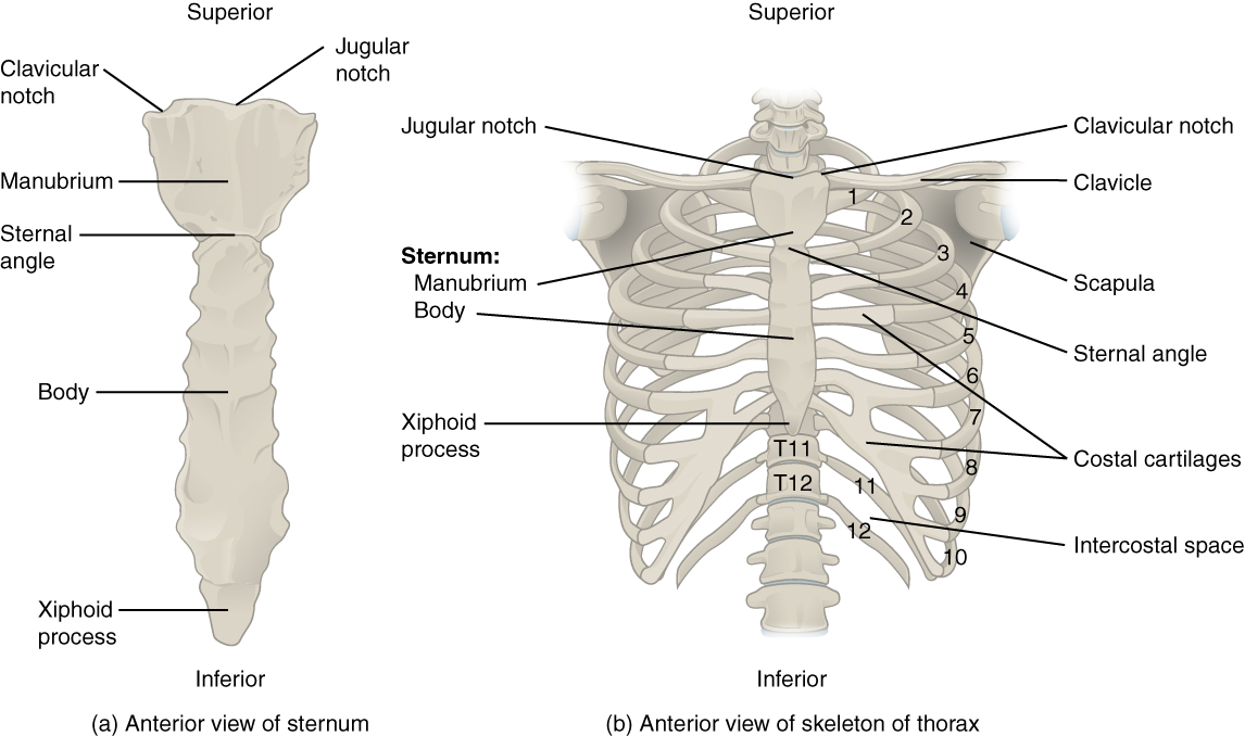
7.4 The Thoracic Cage - Anatomy and Physiology from opentextbc.ca Vintage anatomical drawing medical illustration , pelvis , hip skeleton book page , paper ephemera human anatomy. Bones are not inanimate rock like structures in the human body; A typical long bone shows the gross anatomical characteristics of bone. The image is available for download in high resolution quality up to 4096x4096. The first seven are connected behind with the vertebral column. Long bones, short bones, and flat bones. + benefits of wire mounted bone models: Vertebrae in the thoracic spine on the other hand are less mobile due to being joined by the ribs to the sternum.
Bones are not inanimate rock like structures in the human body;
Bones are not inanimate rock like structures in the human body; The sacrum and coccyx are comprised of. Your rib cage, for example, acts like a shield around your chest to protect important organs inside such as your lungs and heart. Vintage anatomical drawing medical illustration , pelvis , hip skeleton book page , paper ephemera human anatomy. The wider section at each end of the bone is called the epiphysis (plural the two layers of compact bone and the interior spongy bone work together to protect the internal organs. Human body anatomy, body silhouette. The ribs are curved, flat bones which form the majority of the thoracic cage. From a rescued book falling apart at its bindings comes this fascinating vintage medical illustration of the bones of the pelvis and hip. In vertebrate anatomy, ribs (latin: The ribs also bend a little as we breathe; In adults, the cut section would show cancellous bone eighty percent of the skeleton is composed of cortical bones whereas 20% is cancellous in human adults. The hand bones are also known as carpel bones. Have you ever seen fossil remains of dinosaur and ancient human bones in textbooks, television, or in person at a museum?
The vertebrae when we lift heavy weights; human bone anatomy. Head (caput costae) neck (collum costae) body with the upper ribs, closer to the nodule (and in the case of lower ribs, a little further from the nodule) they are curved and have a rough surface that.

